The heart's own arteries
The heart muscle is too thick to feed on the blood inside its chambers. Two arteries, the right and the left coronary, leave the aorta just above the aortic valve as the first branches of the systemic circulation. They run in the grooves on the heart's surface and send branches into the muscle.
The left coronary divides after one or two centimetres. Its anterior interventricular branch runs down the front between the two ventricles, and its circumflex branch curves round the left side to the back. Between them they feed most of the left ventricle.
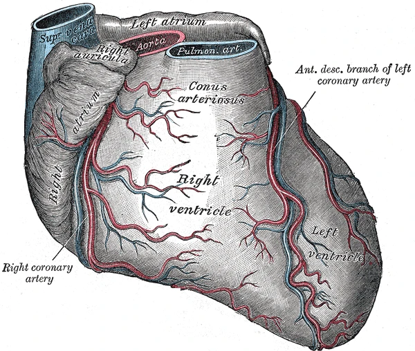 The heart from the front with the right coronary artery and the anterior descending branch of the left coronary artery, Gray's Anatomy, 1918, figure 492Wikimedia Commons, Henry Vandyke Carter, Public domain (Gray492.png)
The coronaries fill mostly between beats, when the muscle relaxes and stops squeezing them. At rest they carry about 4 percent of the heart's output. A heart attack happens when a clot blocks one of them. The muscle beyond the clot gets no oxygen, and after about 20 minutes without blood it begins to die.
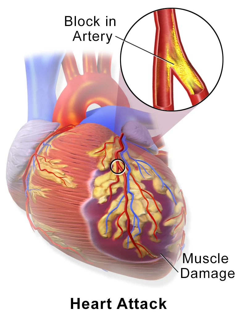 A heart attack: a clot blocks a branch of the left coronary artery and the muscle beyond it is damagedWikimedia Commons, Blausen Medical Communications, CC BY 3.0 (Blausen 0463 HeartAttack.png)
Sources
- Susan Standring (ed.), Gray's Anatomy, 42nd ed. (2020), chapter on the heart.
- Arthur Guyton and John Hall, Textbook of Medical Physiology, 13th ed. (2016), ch. 21.
Prev: The valvesNext: The heart's veins