The aorta
The aorta starts as the ascending aorta, which leaves the left ventricle upwards, about 25 millimetres across. It bends over the heart in the arch, runs down the back of the chest beside the spine as the thoracic aorta, and passes through the diaphragm to become the abdominal aorta.
At the level of the fourth lumbar vertebra it divides into the two common iliac arteries. From the valve to this fork it is about 40 centimetres long.
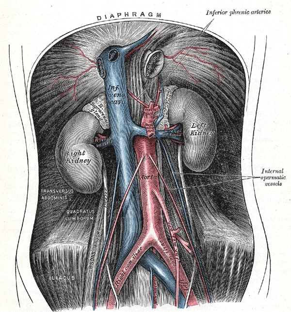 The abdominal aorta and the inferior vena cava, with the kidneys and the fork of the aorta into the common iliac arteries, Gray's Anatomy, 1918, figure 531Wikimedia Commons, Henry Vandyke Carter, Public domain (Gray531.png)
Its wall is elastic. It stretches with every stroke of the ventricle and recoils between strokes, which keeps the blood flowing while the heart refills. With age the wall stiffens, and the pressure the heart has to push against rises.
Sources
- Susan Standring (ed.), Gray's Anatomy, 42nd ed. (2020), chapters on the great vessels and the posterior abdominal wall.
- Arthur Guyton and John Hall, Textbook of Medical Physiology, 13th ed. (2016), ch. 15.
Prev: Air and bloodNext: Head and arms