The valves
Four valves keep the blood moving one way. One valve lies between each atrium and its ventricle. The tricuspid on the right has three leaflets, the mitral on the left has two. Cords tie the leaflets to small muscles in the ventricle wall, so they cannot turn back into the atrium when the ventricle squeezes.
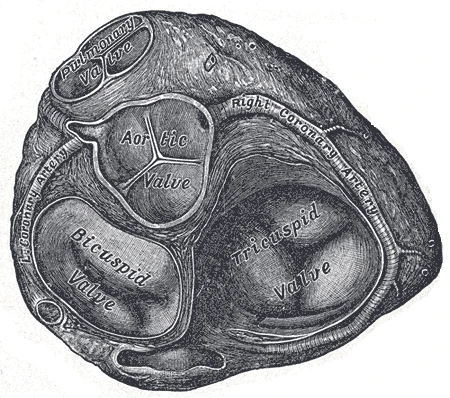 The base of the ventricles with the atria removed: the pulmonary, aortic, mitral (bicuspid) and tricuspid valves, Gray's Anatomy, 1918, figure 495Wikimedia Commons, Henry Vandyke Carter, Public domain (Gray495.png)
At the outlet of each ventricle sits another, the pulmonary valve on the right and the aortic valve on the left. Each has three cusps shaped like pockets, which fill and close as soon as blood starts to flow back.
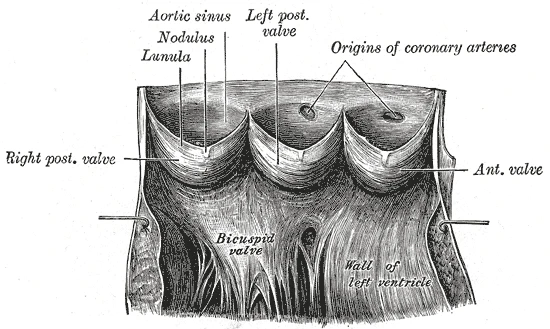 The aortic valve laid open: three cusps shaped like pockets, with the openings of the coronary arteries just above them, Gray's Anatomy, 1918, figure 497Wikimedia Commons, Henry Vandyke Carter, Public domain (Gray497.png)
Through a stethoscope each beat sounds as two. The first sound is the tricuspid and mitral valves closing as the ventricles start to contract, the second the pulmonary and aortic valves closing as they relax. A murmur between them is blood forced through a valve that has narrowed or leaks.
Sources
- Susan Standring (ed.), Gray's Anatomy, 42nd ed. (2020), chapter on the heart.
- Arthur Guyton and John Hall, Textbook of Medical Physiology, 13th ed. (2016), ch. 9 and 23.
Prev: The beatNext: The heart's own arteries