Folds and villi
Inside, the jejunum is lined with circular folds up to 8 mm tall. The folds are covered with villi, finger-like projections about a millimetre long, and each cell on a villus carries about a thousand microvilli on its surface. Each step multiplies the surface that touches the food.
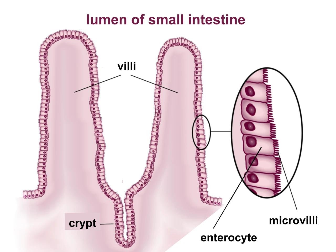 Villi of the small intestine and the microvilli on the surface of their cellsWikimedia Commons, BallenaBlanca, CC BY-SA 4.0 (Villi & microvilli of small intestine.svg)
Older textbooks put the inner surface of the small intestine at 250 square metres, a tennis court. Measurements published in 2014 give about 30 square metres, half a badminton court and still some fifteen times the skin. The enzymes that finish digestion sit on the microvilli, and the lining is renewed about every five days.
Each villus holds a net of capillaries and a lymph vessel, the lacteal. Sugars and amino acids enter the capillaries and go to the liver. Fats are packed into droplets that enter the lacteal and reach the blood by way of the lymph.
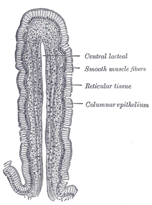 A villus cut lengthwise, with its central lacteal, from a dog's small intestine, Gray's Anatomy, 1918, figure 1059Wikimedia Commons, Henry Vandyke Carter, Public domain (Gray1059.png)
Sources
- Arthur Guyton and John Hall, Textbook of Medical Physiology, 13th ed. (2016), ch. 66.
- Susan Standring (ed.), Gray's Anatomy, 42nd ed. (2020), chapter on the small intestine.
- Herbert Helander and Lars Fändriks, Surface area of the digestive tract revisited, Scandinavian Journal of Gastroenterology 49 (2014): 681 to 689.
Prev: Six metres of gutNext: The mesentery