A second network
Beside the blood vessels runs a second network. It carries a clear fluid, lymph, one way only: from the tissues back to the blood. Its vessels begin as closed tubes in almost every tissue, pass through hundreds of filters, the lymph nodes, and join into the thoracic duct, which ends in the great veins of the neck.
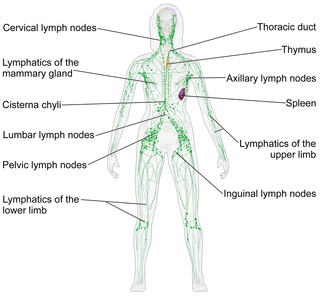 The lymphatic system of a woman: the lymph vessels of the limbs and the breast, the cervical, axillary, lumbar, pelvic and inguinal nodes, the cisterna chyli, the thoracic duct, the thymus and the spleenWikimedia Commons, BruceBlaus (Blausen.com staff), CC BY 3.0 (Blausen 0623 LymphaticSystem Female.png)
The same system holds the organs where white blood cells are made, trained and stored: the red marrow inside the bones, the thymus behind the breastbone and the spleen under the left ribs. Its two jobs, draining fluid and fighting infection, share one set of vessels.
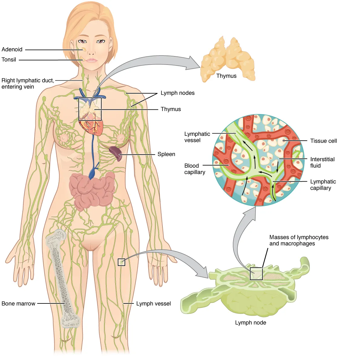 The lymphatic system with its organs, the adenoid, tonsil, thymus, spleen and bone marrow, and enlarged, the lymph capillaries in a tissue and a lymph node, OpenStax, Anatomy and PhysiologyWikimedia Commons, OpenStax College, CC BY 3.0 (2201 Anatomy of the Lymphatic System.jpg)
The nodes and ducts on the model are drawn. The nodes stand where an anatomist placed them in the Z-Anatomy atlas, and the ducts follow the course the anatomy texts give. Their shapes are simplified, and real nodes vary in size and number from one person to the next.
Sources
- Susan Standring (ed.), Gray's Anatomy, 42nd ed. (2020), ch. 4 and 6.
- Arthur Guyton and John Hall, Textbook of Medical Physiology, 13th ed. (2016), ch. 16.
Next: Leaving the capillaries