Inside a node
A lymph node is a bean-shaped capsule, a few millimetres to two centimetres long. Several vessels bring lymph into its rounded side. Inside, the lymph seeps through channels lined with macrophages, which swallow bacteria, dust and debris. It leaves by one or two vessels at the hilum, the dent of the bean, where an artery and vein enter.
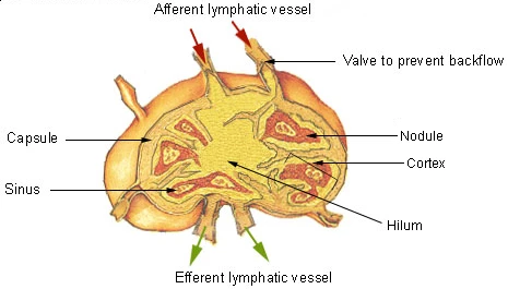 A lymph node cut open: afferent vessels with valves enter its rounded side, the lymph passes the nodules of the cortex and leaves by efferent vessels at the hilum, SEER Training, U.S. National Cancer InstituteWikimedia Commons, SEER, U.S. National Cancer Institute, Public domain (Illu lymph node structure.png)
The outer layer, the cortex, holds round follicles of B cells. Deeper lies a zone of T cells, and in the middle, the medulla, plasma cells release antibodies into the outgoing lymph. Lymphocytes enter the node from the blood through the walls of its small veins, so the cells and the lymph meet here.
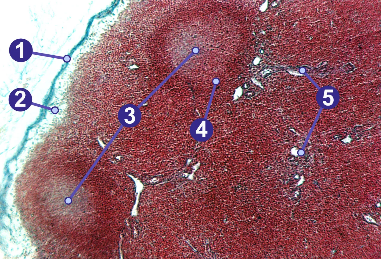 A pig's lymph node under the microscope: 1 capsule, 2 subcapsular sinus, 3 germinal centres, 4 lymphoid nodule, 5 trabeculaeWikimedia Commons, Gleiberg, CC BY-SA 2.0 de (Lymphknoten (Schwein).jpg)
The cut on the model passes through the top of the thigh and the inguinal nodes. The deep ones lie against the inner side of the femoral vein, beside the femoral artery.
Sources
- Susan Standring (ed.), Gray's Anatomy, 42nd ed. (2020), ch. 4 and chapter on the pelvic girdle, hip, gluteal region and thigh.
- Arthur Guyton and John Hall, Textbook of Medical Physiology, 13th ed. (2016), ch. 34 and 35.
Prev: Where the nodes gatherNext: The neck