The first lymph vessels
Lymph vessels begin as closed tubes among the cells, a little wider than blood capillaries. Their wall is a single layer of thin cells that overlap at the edges. Fluid pressing from outside pushes an edge inwards like a flap and enters. Pressure from inside presses the edges together, so lymph cannot leak back out.
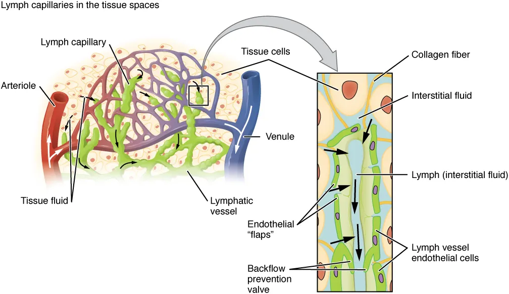 A lymph capillary in the tissue spaces and, enlarged, its wall: overlapping endothelial flaps let tissue fluid in and close against backflow, and collagen fibres anchor the wall, OpenStax, Anatomy and PhysiologyWikimedia Commons, OpenStax College, CC BY 3.0 (2202 Lymphatic Capillaries.jpg)
Fine threads anchor the wall to the tissue around it. When the tissue swells, they pull the tube open and drainage speeds up. Once inside, the fluid is called lymph. It is the same fluid that bathes the tissues, and it carries water, salts, some protein and anything the tissue has shed, such as bacteria or cancer cells.
In the skin of the leg these tubes join into vessels that climb beside the superficial veins, such as the great saphenous vein, to the nodes of the groin.
Sources
- Arthur Guyton and John Hall, Textbook of Medical Physiology, 13th ed. (2016), ch. 16.
- Susan Standring (ed.), Gray's Anatomy, 42nd ed. (2020), ch. 6.
Prev: Leaving the capillariesNext: Valves and pumps