Milky lymph
The lining of the small intestine is covered with villi, folds like fingers about a millimetre tall. Each holds a net of blood capillaries and, in its centre, one lymph vessel, the lacteal. Sugars and amino acids enter the blood and go to the liver by the portal vein.
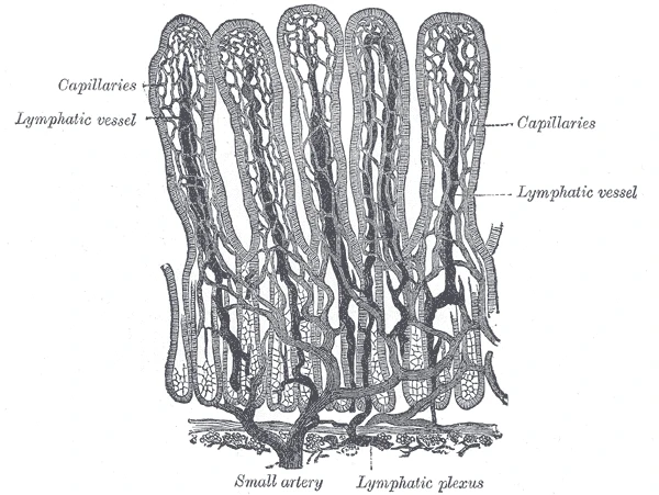 Villi of the small intestine with their blood capillaries and, in the middle of each, a lymph vessel, Gray's Anatomy, 1918, figure 1061Wikimedia Commons, Henry Vandyke Carter, Public domain (Gray1061.png)
Fat takes the other road. The gut cells pack it into chylomicrons, droplets of fat wrapped in protein, too large to enter the blood capillaries, and these slip into the lacteals instead. After a fatty meal the lymph of the jejunum turns milky white and is called chyle.
Chyle flows through the mesentery and its nodes to the cisterna chyli, up the thoracic duct and into the blood in the neck. So most of the fat we absorb reaches the heart and the body before it reaches the liver.
Sources
- Arthur Guyton and John Hall, Textbook of Medical Physiology, 13th ed. (2016), ch. 66.
- Susan Standring (ed.), Gray's Anatomy, 42nd ed. (2020), chapter on the small intestine.
Prev: Peyer's patchesNext: Swelling