1677
Peyer's patches
In the wall of the ileum, the last part of the small intestine, lie oval patches of lymphoid tissue a centimetre or more long, on the side opposite the mesentery. Johann Conrad Peyer described them in 1677. Each patch is a cluster of follicles like those of a node, set just under the lining of the gut.
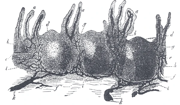 A Peyer's patch in section: villi above, round lymph nodules below, and the lymph vessels injected around them, Gray's Anatomy, 1918, figure 1063Wikimedia Commons, Henry Vandyke Carter, Public domain (Gray1063.png)
Special cells in the lining over a patch take up samples of what passes in the gut and hand them to the lymphocytes beneath. The B cells there make IgA, an antibody that is secreted into the gut and binds germs before they can attach. The wall of the appendix is packed with the same follicles.
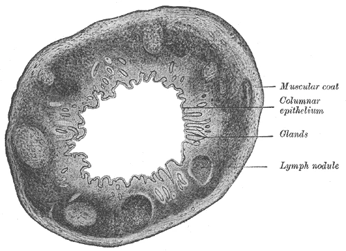 The appendix cut across, with lymph nodules in its wall, Gray's Anatomy, 1918, figure 1074Wikimedia Commons, Henry Vandyke Carter, Public domain (Gray1074.png)
Sources
- Susan Standring (ed.), Gray's Anatomy, 42nd ed. (2020), chapter on the small intestine.
- Johann Conrad Peyer, Exercitatio anatomico-medica de glandulis intestinorum (Schaffhausen, 1677).
- Arthur Guyton and John Hall, Textbook of Medical Physiology, 13th ed. (2016), ch. 35.
Prev: The tonsilsNext: Milky lymph