A school for T cells
Each lobe of the thymus is split into lobules, each with a dark outer cortex packed with young T cells and a paler core, the medulla. Young cells arrive from the marrow. Each builds a receptor by joining gene pieces at random, so every cell recognizes a different shape, and most of those shapes are useless or dangerous.
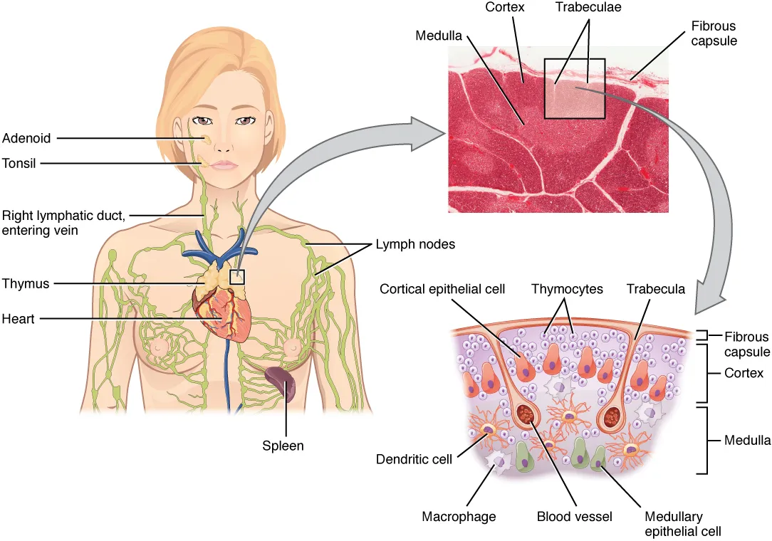 The thymus behind the breastbone, a micrograph of its cortex and medulla, and a diagram of the young T cells among the epithelial cells, OpenStax, Anatomy and PhysiologyWikimedia Commons, OpenStax College, CC BY 3.0 (2206 The Location Structure and Histology of the Thymus.jpg)
The thymus tests them. A cell whose receptor cannot grip the body's own marker molecules dies in the cortex. A cell that grips the body's own proteins too hard dies in the medulla, since it would attack the body. More than nine in ten die there, and the rest enter the blood as mature T cells.
The cut on the model passes through both lobes. Under the microscope the medulla shows round whorls of flattened cells, Hassall's corpuscles, found only in the thymus.
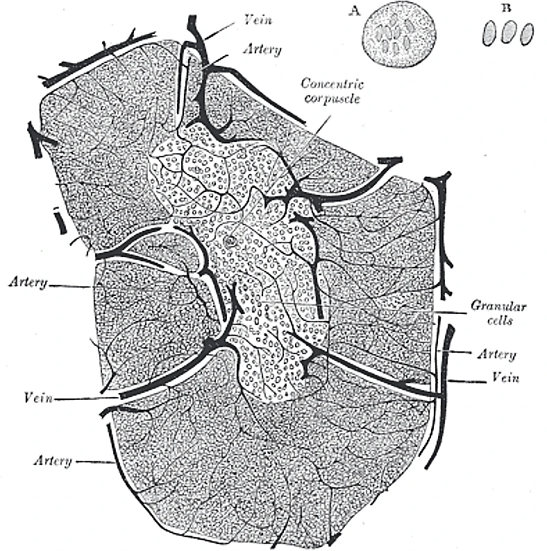 A lobule of the thymus, slightly diagrammatic: a dark cortex round a pale medulla that holds a concentric corpuscle, Gray's Anatomy, 1918, figure 1179Wikimedia Commons, Henry Vandyke Carter, Public domain (Gray1179.png)
Sources
- Susan Standring (ed.), Gray's Anatomy, 42nd ed. (2020), ch. 4.
- Arthur Guyton and John Hall, Textbook of Medical Physiology, 13th ed. (2016), ch. 35.
Prev: The thymusNext: Born in the marrow