Breathing
The diaphragm is the main muscle of breathing. It is a dome of muscle around a central tendon that closes the bottom of the chest. Its fibres start on the lower ribs, the sternum and the lumbar vertebrae. When they contract the dome flattens and moves down, the chest grows taller and air flows into the lungs.
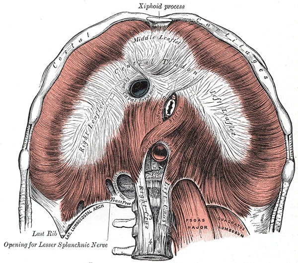 The diaphragm from below, its muscle arranged round the central tendon, with the openings for the inferior vena cava, the esophagus and the aorta, Gray's Anatomy, 1918, figure 391Wikimedia Commons, Henry Vandyke Carter, Public domain (Gray391.png)
Quiet breathing, about 12 breaths a minute at rest, is done almost entirely by the diaphragm. Breathing out is passive: the diaphragm relaxes and the elastic lungs and chest wall spring back. The diaphragm is supplied by the phrenic nerve, from the third to fifth cervical segments of the spinal cord.
Between the ribs lie two main layers of intercostal muscles. The external intercostals lift the ribs and widen the chest in a deep breath. The internal intercostals pull the ribs down when breathing out is forced, as in coughing or blowing out candles, helped by the muscles of the abdomen.
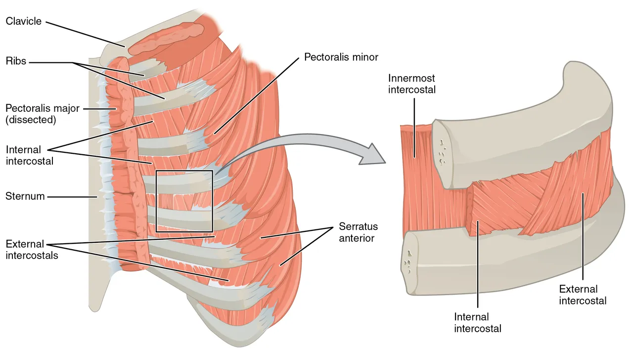 The muscles of the chest wall, with the external, internal and innermost intercostals between the ribs, OpenStax Anatomy and PhysiologyWikimedia Commons, OpenStax, CC BY 4.0 (1114 Thorax.jpg)
Sources
- Susan Standring (ed.), Gray's Anatomy, 42nd ed. (2020), chapters on the diaphragm and phrenic nerve and on the chest wall.
- Arthur Guyton and John Hall, Textbook of Medical Physiology, 13th ed. (2016), ch. 38.
Prev: The chestNext: The abdominal wall