1954
The sliding filaments
By the early 1950s electron microscopes and X-ray studies had shown that a myofibril holds two sets of filaments, thick ones of myosin and thin ones of actin. How they made a muscle shorten was not known. Many researchers thought the long protein molecules folded or coiled up, as a spring does.
On 22 May 1954 the journal Nature printed two papers side by side. Andrew Huxley and Rolf Niedergerke had watched living frog muscle fibres through a new interference microscope. Hugh Huxley and Jean Hanson had studied myofibrils from the psoas muscle of rabbits under a phase-contrast microscope. The two Huxleys were not related and had worked apart.
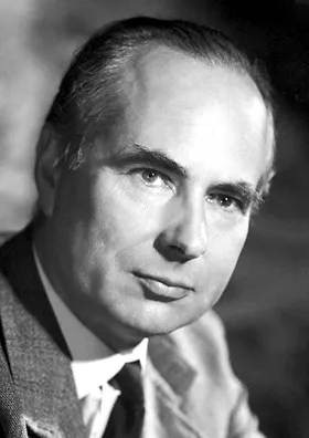 Andrew Huxley, who shared the Nobel Prize in Physiology or Medicine in 1963 for his work on the nerve impulseWikimedia Commons, Nobel Foundation, Public domain (Andrew Fielding Huxley nobel.jpg)
Both teams found that as the muscle shortened, the dark A bands kept their length while the light I bands narrowed. The filaments kept their length and slid past each other. In 1957 Andrew Huxley proposed that heads on the myosin pull the actin along in repeated cycles, the cross-bridges described earlier in this story.
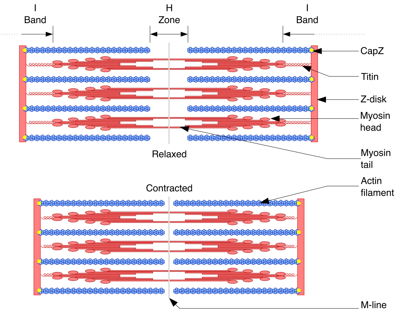 A sarcomere relaxed and contracted, the I bands and the H zone narrowing as the thin filaments slide between the thick onesWikimedia Commons, David Richfield, CC BY-SA 3.0 (Sarcomere.svg), white background added
Sources
- A. F. Huxley and R. Niedergerke, Structural changes in muscle during contraction, Nature 173 (1954): 971-973.
- H. E. Huxley and J. Hanson, Changes in the cross-striations of muscle during contraction and stretch and their structural interpretation, Nature 173 (1954): 973-976.
- A. F. Huxley, Muscle structure and theories of contraction, Progress in Biophysics and Biophysical Chemistry 7 (1957): 255-318.
Prev: Duchenne's faces