Golgi and Cajal
In 1873 Camillo Golgi, a doctor at a hospital near Milan, hardened brain tissue in potassium dichromate and soaked it in silver nitrate. A few nerve cells in every hundred turned black down to their finest branches, while the rest stayed clear. For the first time a whole neuron could be seen. Golgi thought the fibres fused into one net.
From 1888 Santiago Ramón y Cajal, in Barcelona, used the stain on the cerebellum and the cortex of young animals and drew what he saw. Each neuron ended free, close to the next one without joining it, and signals ran one way, from the dendrites through the cell body to the axon.
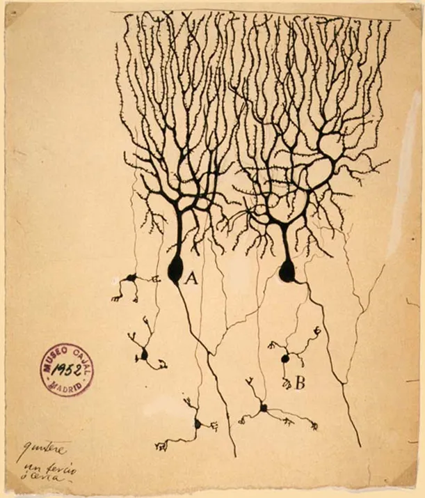 Purkinje cells (A) and granule cells (B) from the cerebellum of a pigeon, drawn by Santiago Ramón y CajalWikimedia Commons, Santiago Ramón y Cajal, Public domain (PurkinjeCell.jpg)
The two shared the Nobel Prize in 1906, and in his lecture Golgi still defended his net. The electron microscope settled the question in the 1950s by showing a gap of a few hundredths of a micrometre at every synapse.
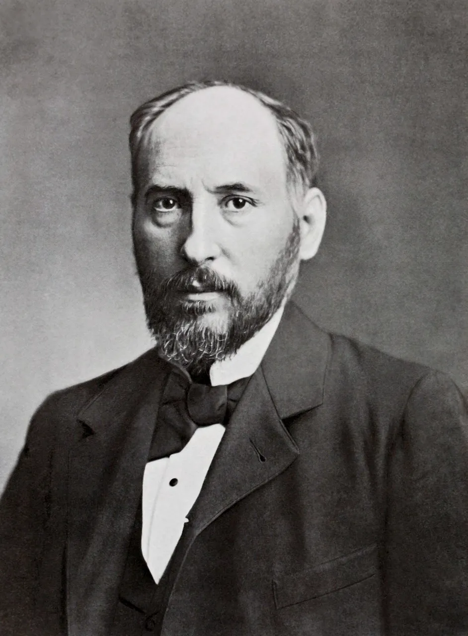 Santiago Ramón y Cajal, in a portrait published by Clark University in 1899Wikimedia Commons, anonymous photographer, restored by Garrondo, Public domain (Santiago Ramón y Cajal (1852-1934) portrait (restored).jpg)
Sources
- Camillo Golgi, Sulla struttura della sostanza grigia del cervello, Gazzetta Medica Italiana, Lombardia 33 (1873): 244-246.
- Santiago Ramón y Cajal, Estructura de los centros nerviosos de las aves, Revista Trimestral de Histología Normal y Patológica 1 (1888): 1-10.
- Camillo Golgi, The neuron doctrine: theory and facts, Nobel lecture, 11 December 1906.
Prev: Broca's patient