The knee jerk
A tap just below the kneecap stretches the tendon of the quadriceps, the muscle on the front of the thigh. Stretch sensors inside the muscle, the muscle spindles, fire. Their fibres run up the femoral nerve to the cord, which they enter at the second to fourth lumbar segments.
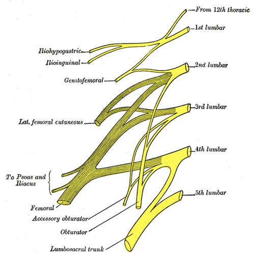 Plan of the lumbar plexus: the femoral nerve forms from the second, third and fourth lumbar nerves, Gray's Anatomy, 20th edition, 1918, figure 822Wikimedia Commons, Henry Vandyke Carter, Public domain (Gray822.png)
There each sensory fibre ends directly on the motor neurons of the same muscle. After that one synapse the command runs straight back out, the quadriceps contracts and the leg kicks. With one tap a doctor tests the nerve, the cord segments and the motor neurons.
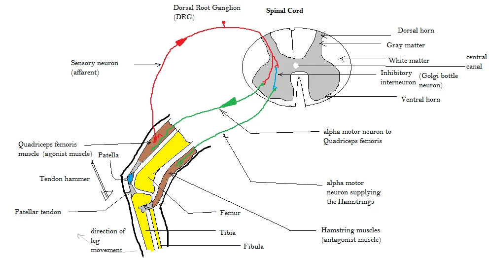 The knee jerk: the sensory fibre from the quadriceps enters the cord through the dorsal root ganglion and excites the motor neuron of the same muscleWikimedia Commons, Amiya Sarkar, CC BY-SA 4.0 (Patellar tendon reflex arc.png)
Wilhelm Erb and Carl Westphal both described the reflex in 1875, in the same issue of a Berlin journal.
Sources
- Arthur Guyton and John Hall, Textbook of Medical Physiology, 13th ed. (2016), ch. 55.
- Susan Standring (ed.), Gray's Anatomy, 42nd ed. (2020), chapter on the internal organization of the spinal cord.
- Wilhelm Erb, Über Sehnenreflexe bei Gesunden und bei Rückenmarkskranken, and Carl Westphal, Über einige Bewegungs-Erscheinungen an gelähmten Gliedern, Archiv für Psychiatrie und Nervenkrankheiten 5 (1875).
Prev: Where the cord endsNext: The brachial plexus