The cord and its nerves
The spinal cord runs down a canal through the vertebrae, from the base of the skull to the small of the back. It is about 45 centimetres long and as thick as a finger, and like the brain it is wrapped in three membranes. Cut across, it shows a butterfly of grey matter inside the white.
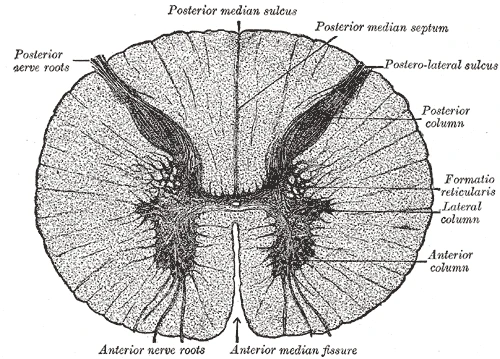 Cross-section of the spinal cord in the chest: a butterfly of grey matter inside the white, with the posterior and anterior nerve roots, Gray's Anatomy, 20th edition, 1918, figure 664Wikimedia Commons, Henry Vandyke Carter, Public domain (Gray664.png)
Between each pair of vertebrae a nerve leaves on each side, 31 pairs in all: 8 cervical, 12 thoracic, 5 lumbar, 5 sacral and 1 coccygeal. Each nerve joins two roots. The back root, with its swelling, the spinal ganglion, brings sensation in. The front root sends commands out. Charles Bell and François Magendie showed this between 1811 and 1822.
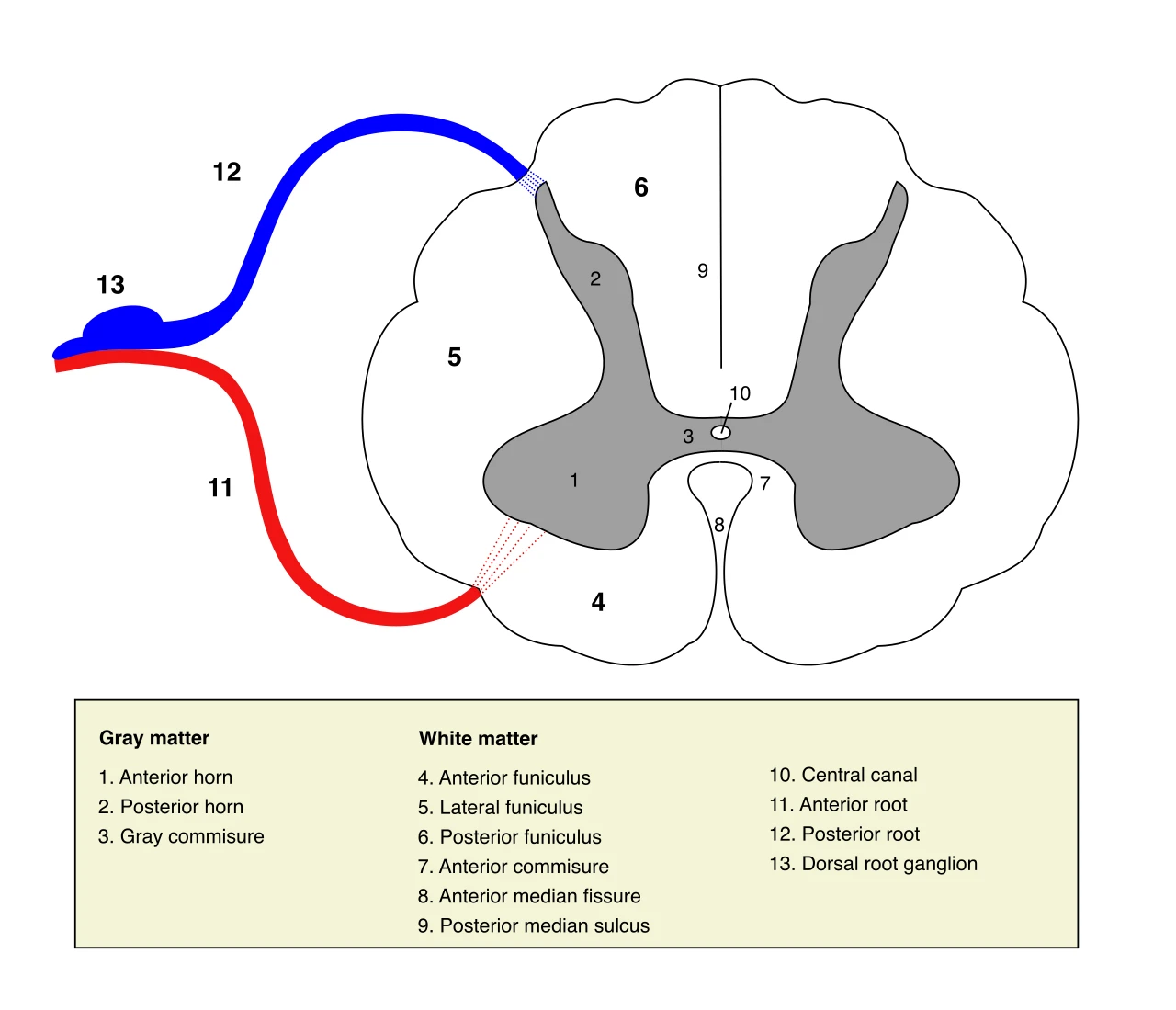 A segment of the spinal cord with its two roots: the posterior root and its ganglion bring sensation in, the anterior root sends commands outWikimedia Commons, Polarlys, CC BY 2.5 (Medulla spinalis - Section - English.svg)
The third, fourth and fifth cervical nerves form the phrenic nerve to the diaphragm. An injury to the cord above the third cervical segment stops breathing.
Sources
- Susan Standring (ed.), Gray's Anatomy, 42nd ed. (2020), chapter on the gross anatomy of the spinal cord and spinal nerves.
- Susan Standring (ed.), Gray's Anatomy, 42nd ed. (2020), chapter on the internal organization of the spinal cord.
Prev: From cortex to cordNext: Where the cord ends