The arches of the foot
The bones of the foot form arches. The long arch on the inner side, seen here, runs from the heel through the talus and the navicular to the first three metatarsals. A lower arch runs along the outer side, and a third arch crosses the foot. Standing, the weight rests on the heel and the ball of the foot.
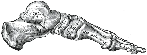 The skeleton of the foot from the inner side, with the arch from the calcaneus to the first metatarsal, Gray's Anatomy, 1918, figure 290Wikimedia Commons, Henry Vandyke Carter, Public domain (Gray290.png)
Bone shapes, ligaments and muscles hold the arches up. The spring ligament runs from the calcaneus to the navicular and carries the head of the talus. The long plantar ligament ties the calcaneus to the cuboid and the metatarsals. Under them the plantar fascia, a thick band, ties the heel to the toes like a bowstring.
At each step the arch flattens a little as the weight comes on and stiffens as the heel lifts. In a flat foot the inner arch is low or missing, which in most people causes no trouble.
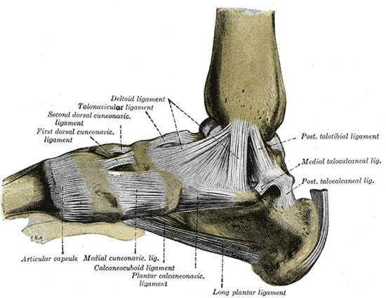 The ligaments on the inner side of the foot, with the plantar calcaneonavicular (spring) ligament and the long plantar ligament, Gray's Anatomy, 1918, figure 354Wikimedia Commons, Henry Vandyke Carter, Public domain (Gray354.png)
Sources
- Susan Standring (ed.), Gray's Anatomy, 42nd ed. (2020), chapter on the ankle and foot.
Prev: 26 bones of the footNext: Three kinds of joint