How bones grow
Most bones begin in the embryo as models of cartilage. From about the seventh week, bone starts to replace the cartilage in the middle of each shaft and spreads toward the ends. The flat bones of the skull roof form in a different way, directly in a sheet of membrane.
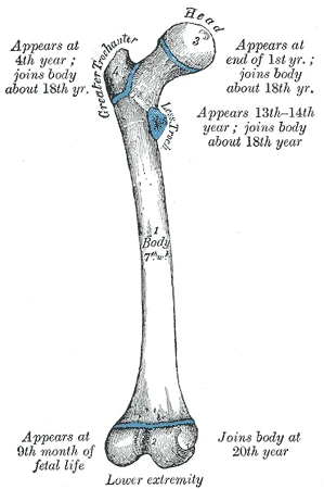 The femur's five centres of bone, with the age at which each appears and joins the shaft, Gray's Anatomy, 1918, figure 252Wikimedia Commons, Henry Vandyke Carter, Public domain (Gray252.png)
Later, bone also starts to form in the ends. Between each end and the shaft a thin disc of cartilage stays, the growth plate. Its cells divide and bone replaces them on the shaft side, so the bone lengthens. The plates at the knee ends of the femur and the tibia add most of the leg's length.
At the end of puberty, a few years earlier in girls than in boys, the plates turn to bone and growth in height stops. The last plate to close, at the inner end of the clavicle, closes in the mid twenties.
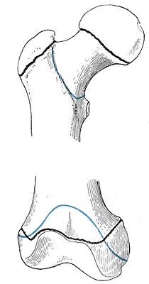 The ends of a young adult's femur, with the lines where the growth plates closed in black, Gray's Anatomy, 1918, figure 253Wikimedia Commons, Henry Vandyke Carter, Public domain (Gray253.png)
Sources
- Susan Standring (ed.), Gray's Anatomy, 42nd ed. (2020), chapter on bone and cartilage.
- J. Gordon Betts and others, Anatomy and Physiology (Houston: OpenStax, 2013), section 6.4.
Prev: Ball, hinge and pivotNext: A broken bone heals