Inside a synovial joint
In a synovial joint the bone ends are capped with smooth cartilage a few millimetres thick and closed in a capsule. The capsule is lined by the synovial membrane, which makes synovial fluid, a slippery liquid like egg white. The fluid lets the cartilage slide and feeds it, since cartilage has no blood vessels.
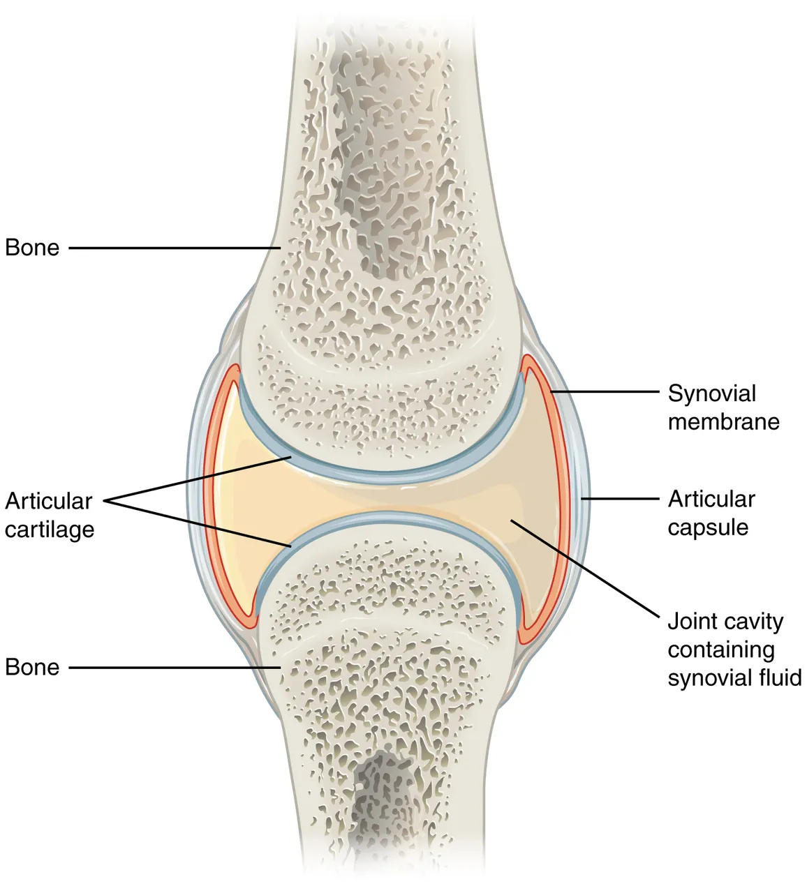 A synovial joint: articular cartilage on the bone ends, the joint cavity with synovial fluid, the synovial membrane and the capsuleWikimedia Commons, OpenStax College, CC BY 3.0 (907 Synovial Joints.jpg)
The hip is a deep synovial joint, shown here by its lit capsule. The head of the femur sits in the acetabulum of the hip bone, deepened by a rim of fibrous cartilage, the labrum. A small ligament runs from the head to the floor of the socket.
Ligaments thicken the capsule. The iliofemoral ligament across the front of the hip is one of the strongest in the body. Standing, it is pulled tight and stops the hip bending backward.
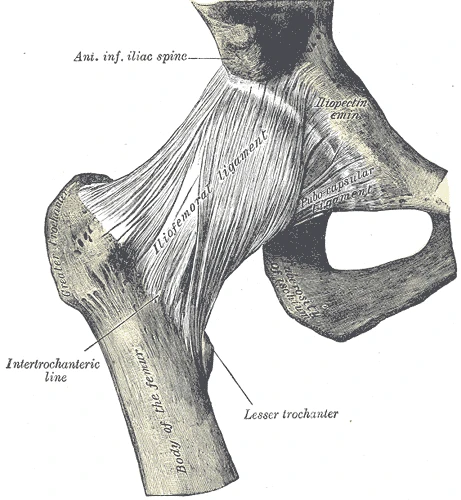 The right hip joint from the front, with the iliofemoral ligament across the capsule, Gray's Anatomy, 1918, figure 339Wikimedia Commons, Henry Vandyke Carter, Public domain (Gray339.png)
Sources
- Susan Standring (ed.), Gray's Anatomy, 42nd ed. (2020), chapters on the functional anatomy of the musculoskeletal system and on the hip.
- J. Gordon Betts and others, Anatomy and Physiology (Houston: OpenStax, 2013), section 9.4.
Prev: Three kinds of jointNext: Ball, hinge and pivot